As in Incident command...

IC established!
We'll have several different sections reporting in - recent research, local topics, or highlighting areas of the Sponsor Hospital Council of Greater Bridgeport protocols.

*** Keep in mind - this website does not replace your protocols, and these posts do not reflect SHCGB or Bridgeport Hospital policies. This is a place to discuss research, controversies, or discuss possible future protocols. When in doubt, check your current protocols through the official source.

Thursday, May 30, 2013

Atrial Fibrillation: Sure, you can cardiovert it! But should you? (Part 1)

Some cardiac arrhythmias are exciting and fun for the prehospital provider. Ventricular fibrillation has, essentially, only one proven therapy, but needs a well-choreographed team to deliver it. Paroxysmal supraventricular tachycardia, on the other hand, has a far less dire prognosis, but the treatment is generally safe and dramatic - very satisfying for both the patient and provider!

Milwaukee Beers ≠ "well-choreographed team"
Unlike VF or PSVT, however, atrial fibrillation isn’t a fun rhythm to deal with, either in-hospital or prehospital, and there are many ways to screw up. Even the new edition of Nancy Caroline's Emergency Care in the Streets has little to say about management of atrial fibrillation, only mentioning that 
"Prehospital treatment of atrial fibrillation is rare because of the risks involved."
However, they don't describe those risks, or how to avoid them!

They are likely correct about the "rare" part - one study of atrial fibrillation treated by EMS backs this up - you just aren't likely to have a patient with atrial fibrillation that needs cardioversion or emergent diltiazem. On the other hand, atrial fibrillation is really common in EMS, and we should know a bit more about this. There are lots of recent articles and old insights about AF - how about we start with 4 things?

1. AF is often a symptom, not the disease
Some rhythms require treatment, regardless of the history or exam. In other words, sometimes we "treat the monitor."  AF is not like this.

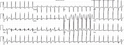
Example: A medic recently brought us an older female with palpitations. Evidently this had started about 3 hours prior, and was just getting worse. She had been taking all her "heart pills," but had had some vomiting and diarrhea for the past day and a half. Her HR was 180, and she was somewhat hypotensive at 95/60. Her sat and RR were basically normal. 

A 12-lead was obtained:

ST-segment changes, rapid AF and hypotension - should the patient receive diliazem, metoprolol, or even cardiovert? These can the right answers for many rhythms with & without hypotension (e.g. VT, PSVT), but AF can be different.

The medic decided, based on the recent history of volume losses, that a fluid bolus should be tried. After 300 ml of NS he saw a change in heart rate, and recorded ECG #2:


The blood pressure settled out at 120s/70s, and the rest of transport was uneventful. She ended up getting some diltiazem in the ED, but it looked more like dehydration that caused her tachycardia, as well as impaired absorption of her medications.

This is very common - AF is often provoked or worsened by non-cardiac problems. A drinking binge can do it, and so can hyperthyroidism. A low magnesium, in some cases, may be responsible. Both a large pulmonary embolus (because of the right atrial strain), and an MI, may kick off AF. And as for alcohol...

2. Alcohol (too much or too little) and AF.
He was weak, had vomited at least 10 times that day, and couldn't stop shaking. This 40 y.o. man had stopped drinking 2 days ago, and had not done well since. The palpitations were the final straw, pushing him to come to the ED.

"Excuse me, did something crawl down your throat and die"
"It didn't die!"
His heart rate was bumping up over 200, but the ECG caught him at a relatively slow point:


His labs confirmed that he has dehydrated, with low levels of magnesium and potassium. His alcohol history, both the excess, and the abrupt cessation, also likely contributed to provoking a new-onset atrial fibrillation. 

He clearly didn't need cardioversion, but it also seemed premature to use diltiazem or metoprolol first. He received a liter of saline, some magnesium and potassium, as well as Valium for the withdrawal. This went a long way towards improving his heart rate, and he only needed a small dose of metoprolol after all that.

So, while he had atrial fibrillation, he had other medical issues (like the withdrawal) that were more important. This is actually pretty common - about 75% of patients who come to the emergency department who have AF on their ECG actually have a different primary diagnosis - the top three diagnoses are CHF, pneumonia, and chest pain

Patients with AF and an Alternative Primary Diagnosis in the ED

3. This is especially true for sepsis and AF.
I recently saw a patient brought in from a nursing home with AF at 170-180 bpm, as well as hypotension. Sorta looked like this:

LITFL
However, she also had a new history of a cough and altered mental status, although no documented fever. I decided to try a liter of saline first, rather than a bolus of diltiazem, thinking that the rapid ventricular response was due to pneumonia and metabolic stress. Fortunately, the bolus dropped the heart rate down to 110-120, and raised her BP. She was admitted to the ICU with a bad case of sepsis and pneumonia, no diltiazem or metoprolol needed.

This case was not unusual - it turns out there is a HUGE relationship between sepsis and AF. Some surprising facts:
AF & sepsis = bad news bears.

It isn't clear how AF should be treated in severe sepsis, besides treating the source of infection and supportive care. The use of rate-controlling medications (diltiazem and metoprolol) may "mask" the signs of sepsis, complicating the use of fluid boluses or pressors. Cardioversion with drugs or electricity pose their own hazards.

So, if your nursing home patient has new AF, think: Could this be sepsis?


To be continued!
So far these patients I've discussed haven't been terribly unstable. In my last example (to be posted soon!) I'll discuss a truly critical patient with paroxysmal AF, where cardioversion was not performed. I've already shown the ECG on the Mill Hill Ave Command Facebook page, but I'll share more elements, and how they relate to management.

Thursday, May 9, 2013

Two ECGs - which goes to the cath lab?

A recent study suggests that a computer interpretation of the ECG can be extremely specific for diagnosing a STEMI; i.e. if the computer reads 

*** ACUTE MI  ***

you can take that to the bank.

This hasn't fit with my experience, and so I was very interested in Peter Canning's latest post, since it validated my suspicions. He found that the ECGs his system are acquiring show surprisingly poor sensitivity and specificity for STEMI, if you simply rely on the computer to diagnose. 

As an illustration of this point, I submit ECGs from 2 patients. 

(For more pairs of ECGs that show the problem with relying on the computer diagnosis of STEMI, click on the label "Paramedics need to read ECGs..." on the right.)

Case 1
Let's say that this was a middle-aged female, who started having substernal chest pain about 15 minutes ago. The EMS 12-lead shows:


Aside from diagnosing the patient as "borderline," anything else look suspicious?

Case 2
Again, a middle-aged female, this time with pleuritic chest pain and wheezing. An ECG obtained 5 minutes after arrival in the ED shows:


What's an appropriate next step? Call in the (cardiology) cavalry, or do a little sleuthing?

Call for a bat-stent? (source)
Resolution
If you look closely at  few of the leads, especially V3, you can see small spikes preceding the QRS. Since the computer hadn't seemed to notice, I adjusted the settings to recognize pacemakers. A second ECG then showed pretty much the same complexes, but a very different interpretation.

Fixed!
She turned out to have a fairly ordinary case of COPD.

How about case #1?

Evidently the patient was first transported to a non-PCI capable hospital. About 2 hours later she was on her way to a different hospital for an urgent cardiac catheterization. This gave EMS a unique opportunity to capture the evolution of the ECG over a time frame that we don't often find in urban/suburban EMS.


Frankly, I'm inclined to agree with the computer this time! But what did the computer "miss" on the first ECG?

Hyperacute T waves
As Peter found after analysis of his system's STEMIs, computers aren't good at recognizing the earliest sign of an MI on an ECG, the hyperacute T-wave. These are transient features, before the ST segment has had a chance to elevate, and EMS is in a unique position to find these on their initial ECG.

Stephen Smith has some great examples, some of which look very similar to case #2 here. For instance, this ECG was acquired by EMS, and was instrumental in suggesting ACS to the emergency physician:

Dr Smith's ECG Blog - 6/2011
Another case involved an anterior MI that was misdiagnosed as hyperkalemia because of the magnitude of the hyperacute T-waves:

Dr Smith's ECG Blog - 2/2009
Very similar to our patient #2!

The Bottom Line
For more teaching on hyperacute T-waves, follow the links above to the blogs written by Peter Canning or Dr Smith, or check out this review.

And remember - sometimes you have to treat the monitor, not just the patient. Just make sure you're not treating a mistaken computer!


Tuesday, May 7, 2013

The Chain of Survival - done right!

The American Heart Association has been using the phrase "chain of survival for years now. This imagery is used to emphasize the importance of an integrated response to a cardiac arrest. This is common sense - you and your gold patch aren't worth much if no one does CPR before you arrive. Or if no one even calls in the first place!


"He'll be okay!" - Source
We recently had a patient brought into the ED which beautifully illustrated this "chain" concept. And while this case did not get onto channel 12, it's a great example of what can be done when all the pieces work they way they should. I'll quote the AHA concept behind each link, and describe how it worked in that case.


1. Immediate recognition of cardiac arrest and activation of the emergency response system 
 Our patient, a 50-ish female, collapsed while at work. This was witnessed by her coworkers, fortunately, and 911 was immediately called.

2. Early CPR with an emphasis on chest compressions
Rather than waiting for EMS to arrive, or even having to be coached by 911 dispatchers, coworkers started CPR.

"They were doing quality CPR when we got there," recalled Christopher Lovell, the AMR paramedic who responded. "They knew what they were doing - probably had a medical response team at the company, from the way they were acting."

3. Rapid defibrillation
Even while the AMR unit was still enroute, the patient's coworkers were giving her every chance. They grabbed the AED available onsite, and they applied the pads, analyzed, and gave shock #1.

4. Effective advanced life support 
When the ALS unit arrived, they found the patient to be in VF, and gave shock #2. The VF continued, and they gave two more defibrillations before converting her into a sinus rhythm. Her blood pressure and oxygen saturation bounced back almost immediately, and they held off on intubating. A bolus of amiodarone was given to prevent VF recurrence, and they grabbed a 12-lead during transport:


Thoughts?

5. Integrated post–cardiac arrest care
EMS arrived in the ED with a patient who had great vitals (indeed, she has hypertensive), but who continued to have a poor mental status. She required RSI for intubation, while therapeutic hypothermia was started with ice bags. Her second ECG looked a lot like the first one:


After a detour for a head CT (She was oddly hypertensive for a post-cardiac arrest patient, raising suspicion that a subarachnoid or intercerebral hemorrhage had triggered the arrest. It happens.), cardiology swooped in to take her to the cath lab.

When I later got a chance to talk with the cardiologist who cathed the patient, he was thrilled to talk about the case. First of all, the infarct-related artery was not a surprise - a 100% occluded RCA.

But more importantly, the therapeutic hypothermia that we had started in the ED wasn't continued, but for all the right reasons. "By the time I had finished deploying the stent, she was moving around. We extubated her in the ICU, and she was talking to us an hour later!"

That's a good reason, we can all agree.

The Bottom Line
It's not just a catchy phrase or a silly graphic. The "chain of survival" works, and it depends on a robust, effective response from each link. 


'Cause even this guy needs the 4 other links!